Review of Interactive Color Atlas of Histology
Last Update 10/21/2017
 |
| Interactive Color Atlas of Histology Student Version 2.0 |
Disclosure: No specific funding received from attributed company for writing this review
As a medical student, it was inevitable to look for med reference in the form of back-aching hundred pages book to graduate gracefully, or instead, despicable. Those books are the manifestation of a sword in the hand of the so-called med student of 20th-century era. The thicker, the sharper, but unfortunately, also heavier. I doubt that most med-student practice bodybuilding, therefore strain and deformity is common.
Nevertheless, technology plays its part relieving the reliever's discomfort. Lippincott Williams & Wilkins, an imprint publisher, developed this one example of histology analyzing practice software. Interactive Color Atlas of Histology is a book that provides CD-Rom containing the mentioned application.
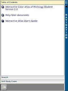
From the welcoming picture, we can see four main panels which consists of Navigation, Slide Tray, Data, and the most important, the Histology Viewer.
1. Navigation

Comprised of three tabs that are "Table of Contents", "Search", and "Self-Study Exam".
Table of Contents
consists of "Interactive Color Atlas of Histology", "Help", and "User's Guide".
- Interactive Color Atlas of Histology:
- The Cell
- Epithelium and Glands
- Connective Tissue
- Cartilage and Bone
- Blood and Hemopoiesis
- Muscle
- Nervous Tissue
- Circulatory System
- Lymphoid Tissue
- Endocrine System
- Integument
- Respiratory System
- Digestive System I-Oral Region
- Digestive System II-Alimentary Canal
- Digestive System III-Digestive Glands
- Urinary System
- Female Reproductive System
- Male Reproductive System
- Special Senses
All of those divisions have further subdivisions in detail, and all are in healthy but no pathological condition.
1. System Requirements:
.
- Help:
1. System Requirements:.
|
IBM-PC or compatible computer that minimally includes:
|
Macintosh PowerPC computer that minimally includes:
|
|
|
|
|
|
|
|
|
|
|
|
|
|
|
2. Technical Support
3. Program Credits
4. Copyrights
5. License Agreement.
This help section provides the necessary components to make sure students will not face any service or reliability issue.
Contains "Getting Started" and all detailed explanations, quite comprehensive.
The search engine will only give me figures which title contain the associated keyword. It's proven when I finally typed "Monkey" and 168 images appear on the view panel.


3. Program Credits
4. Copyrights
5. License Agreement.
This help section provides the necessary components to make sure students will not face any service or reliability issue.
- User's Guide:
Contains "Getting Started" and all detailed explanations, quite comprehensive.Search
Savvy and working search engine that might compete with the penguin algorithm. When I type "Renal", it shows one human paraffin section renal papilla in the viewer panel and other three different plastic section renal images on the slide tray panel. When I type "Kidney", it shows 17 varied section kidney pictures in viewer panel and disparate sum of accompanying pictures on slide tray panel.The search engine will only give me figures which title contain the associated keyword. It's proven when I finally typed "Monkey" and 168 images appear on the view panel.
Self-Study Exam
- USMLE-Style Review Exams
- Basic Tissues Exam
- Organ Systems Exam
- Comprehensive Exam
- Chapter Review Exams
- The Cell
- Epithelium and Glands
- Connective Tissue
- Cartilage and Bone
- Blood and Hemopoiesis
- Muscle
- Nervous Tissue
USMLE-Style is multiple choice while Chapter Review is written and true/false exam. The first mentioned has 96 comprehensive questions whether the later has 569 questions, all based on the figures. Both of them served in study mode and test mode.
Study mode will give you correct answer in each question, test mode will give you score and the correct answer after you finished the exam. It's an elaborate approach in improving student's knowledge.
2. Slide Tray

It's a panel that shows you the rest of the image in a subcategory you open while looking at an object of the contents. Basically the same as the table of content's subsection, just shown in the image form, not a sentence. But this panel also equipped with handy Related Image and History drop-down. The developer could make a histology internet browser of their own if they want to.
3. Data

It gives you the information of the related image viewed. All of the images that shown in the Histology Viewer are complemented with different labels and arrows indicating the landmarked cell or organelle, and, the explanations are served on this panel.
4. Histology Viewer
It's the core of the art where we practice our microscopic vision. The images are shown in the best staining and magnification. Albeit made from human, many are made from monkey. They are available in plastic, paraffin section, smear, and electron microscope.
There exist twelve buttons to accompany the picture:
- Standard mode
- Full-screen mode
- Compare mode
- Thumbnail gallery
- Hot spots on/off
- Help
- Go to Previous
- Go to Next
- Zoom +
- Full zoom in/out
- Zoom -
- All labels on/off
5. All of the images have labels and lines, but they can be replaced with yellow circles in case you want to start memorizing.
12. Moreover, you can remove the labels and the lines too, to see the raw image.
All panel have the expand button. The application I cited is in the 2.0 version, and improvement is highly likely to happen.
In my experience, any easy to learn platform will save my time a lot. All the incoming exam are evolving, and this gadget-friendly app is essential in order to thrive and compete. Based on ease of use and quality content but no pathological image, it's 8/10.
.jpg)

Verry good
ReplyDeleteLiked
ReplyDeleteNice !
ReplyDelete